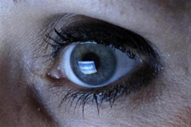
Corneal epithelial stem cells procured from a deceased donor and expanded in the lab were transplanted in the patient’s eye
A study released in STEM CELLS Translational Medicine provides a deeper and clearer understanding of how patients suffering vision loss in both eyes due to limbal stem cell deficiency (LSCD) might benefit from a stem cell transplant using donated cells. This is the first randomised, controlled study to investigate the safety and efficacy of this method, in which corneal epithelial stem cells procured from a deceased donor and expanded in the lab were transplanted in the patient’s eye.
Of the 45 million people worldwide who suffer from bilateral blindness (that is, blindness in both eyes), the World Health Organization estimates more than 20 per cent are due to corneal diseases. LSCD is considered one of the most severe and difficult of these to treat. The limbus, located at the junction of the cornea and sclera, provides a steady source of epithelial stem cells that keep the cornea healthy. If these stem cells are damaged or disrupted – often the result of chemical or thermal trauma, congenital defect or even wearing contact lenses for too long – LSCD might result. The lack of stem cell regeneration leads to severe vision loss, chronic pain and glare, among other symptoms.
Autologous limbal stem cell transplantation, which uses stem cells collected from healthy tissue in the patient’s own eye, has been shown to be effective in treating LSCD when it is confined to one eye but is not practical for treating bilateral disease. Several recent studies have focussed on using allogeneic (donated) material for treating LSCD, but the results are unclear due to several variables in the studies and the fact that none contained a control arm against which results could be compared.
“We wanted to address that in our study,” said co-investigator John Campbell, of the Scottish National Blood Transfusion Service, Edinburgh. “In doing so, we focussed on the manufacture of allogeneic ex vivo expanded corneal epithelial stem cells on amniotic membrane using Good Manufacturing Practice (GMP) standards, as well as the safety and efficacy of this investigational medicinal product (IMP).”
The clinical trial was designed as a randomised, controlled and partially blinded phase I/II trial. “We hypothesised that the use of amniotic membrane, topical autologous serum eye drops and systemic immunosuppression may have therapeutic benefit,” Prof Campbell explained. “By this highly controlled design it was anticipated that the true contribution of stem cells in the IMP would be quantified.”
Patients were divided into groups that received either corneal epithelial stem cells cultured on amniotic membrane (IMP) or control amniotic membrane only. They also received systemic immunosuppression. Of the 16 patients treated, 13 completed all assessments. When the trial ended after 18 months, no adverse reactions were attributed to the IMP itself. Nine of the 13 patients showed improvement in their visual acuity scores, with no statistically significant differences between the IMP and control groups. However, patients in the IMP arm demonstrated significant, sustained improvement in ocular surface score, while those in the control arm did not.
“This trial demonstrates that sustained benefit is achieved by the IMP in ocular surface disease compared with controls. That leads us to believe that this intervention warrants further study in larger sample sizes in a Phase III study,” reported co-investigator Sajjad Ahmad, who during the time of the study was with St Paul’s Eye Unit, Royal Liverpool University Hospital. Professor Baljean Dhillon, Chair of clinical ophthalmology, University of Edinburgh commented, “This is the first RCT in this field and sets the standard for multidisciplinary clinical trial design and delivery of cell-based therapeutics in regenerative ophthalmology.”
New studies would also benefit from concentrating on a single disease group to eliminate some of the variables in this study, the researchers added. “In particular,” Prof Dhillon said, “Our findings suggest some clinical relevance for aniridia patients. This is a genetic condition characterised by a malformed iris that greatly hampers vision. Further studies focussed on how the IMP impacts aniridia-related keratopathy might shed new light on how we can best treat this blinding eye disorder.”
“Lumbar stem cell disease can be a devastating disease leading to sight loss,” said Anthony Atala, , Editor-in-Chief, STEM CELLS Translational Medicine and director of the Wake Forest Institute for Regenerative Medicine. “This trial shows that sight improves and the treatment is safe. We look forward to seeing the long-term outcomes of this technology.”
- Advertisement -


Comments are closed.